

Thursday, September 21, 2023
Wednesday, September 20, 2023
What is canine influenza (dog flu)?
Canine influenza (also known as dog flu) is a contagious respiratory disease in dogs caused by specific Type A influenza viruses known to infect dogs. These are called “canine influenza viruses.” No human infections with canine influenza have ever been reported. There are two different influenza A dog flu viruses: one is an H3N8 virus and the other is an H3N2 virus. Canine influenza A(H3N2) viruses are different from seasonal influenza A(H3N2) viruses that spread annually in people.

Can canine influenza viruses infect people?
In general, canine influenza viruses are thought to pose a low threat to people. To date, there is no evidence of spread of canine influenza viruses from dogs to people and there has not been a single reported case of human infection with a canine influenza virus in the U.S. or worldwide. In 2016, CDC used the Influenza Risk Assessment Tool to evaluate the potential pandemic risk of canine influenza H3N2 viruses and found it to be low.
However, influenza viruses are constantly changing and it is possible that a canine influenza virus could change so that it could infect people and spread easily between people. Human infections with novel (new, non-human) influenza A viruses against which the human population has little immunity are concerning when they occur because of the potential that a pandemic could result. For this reason, the World Health Organization global surveillance system has led to detection of human infections by novel influenza A viruses of animal-origin (such as avian or swine influenza A viruses), but to date, no human infections with canine influenza A viruses have been identified.
Where did canine influenza viruses come from and how long have they been around?
Canine influenza H3N8 viruses originated in horses, spread to dogs, and can now spread between dogs. H3N8 equine influenza (horse flu) viruses have been known to exist in horses for more than 40 years. In 2004, cases of an unknown respiratory illness in dogs (initially greyhounds) were reported in the United States. An investigation showed that this respiratory illness was caused by equine influenza A(H3N8) viruses. Scientists believe this virus jumped species (from horses to dogs) and has adapted to cause illness in dogs and spread among dogs, especially those housed in kennels and shelters. This is now considered a dog-specific, or canine, H3N8 virus. In September 2005, this virus was identified by experts as a “newly emerging pathogen in the dog population” in the United States. It has now been detected in dogs across much of the United States.
Canine influenza H3N2 viruses originated in birds, spread to dogs, and can now spread between dogs. Transmission of H3N2 canine influenza viruses to cats from infected dogs has been reported also. Canine influenza A H3N2 viruses were first detected in dogs in South Korea in 2007, and also have been reported in dogs in China, Thailand, and Canada. H3N2 canine influenza viruses were first detected in the United States in April 2015, and has now been found in more than 30 states. To date, the H3N2 canine viruses reported in the U.S. have been almost genetically identical to canine H3N2 viruses previously reported only in Asia.
How does the issue of canine influenza apply to dogs imported into the US from other countries?
Both of these canine influenza viruses (H3N8 and H3N2), are now considered endemic in dogs in the United States. Additionally, at this time there is no evidence that canine influenza affects humans nor that it has pandemic potential. If there were evidence that canine influenza viruses were able to infect people with the potential for sustained human to human spread, CDC would execute its existing authorities to limit the introduction and/or spread of that pandemic strain either into or within the U.S.
CDC regulations require that dogs be healthy to enter the United States, thus dogs may be denied entry or further evaluated if they look like they are sick with a communicable disease such as canine influenza. CDC can require a veterinary examination at the owner’s expense for dogs that appear sick, or a necropsy (animal autopsy) for dogs that are dead upon arrival in the United States.
Multiple agencies may have regulatory authority over pets such as dogs and cats when they enter the United States. CDC works closely with other federal agencies to protect U.S. borders against diseases in humans that are carried by animals and animal products. The United States Department of Agriculture (USDA) Animal and Plant Health Inspection Service (APHIS) Veterinary Services (VS) has animal health requirements related to bringing (importing) a pet dog to the United States from a foreign country.
What is CDC doing about canine influenza?
Current CDC influenza virus regulations apply only to viruses with pandemic potential in people. However, CDC is doing a number of things to be prepared in the unlikely event that canine influenza becomes a threat to humans or to animals other than canines. First, CDC continues to conduct year-round surveillance for seasonal and novel influenza A viruses and all human infections with novel influenza A viruses are fully investigated. Human infection with a novel influenza A virus of animal origin is reportable to CDC; no human infections with canine influenza viruses have been reported to date. Second, CDC and USDA APHIS VS have existing collaborative protocols to work together in the event of outbreaks of novel influenza A viruses. These same protocols would be followed in the event of an outbreak of canine influenza with suspected human infections. Third, CDC conducted a risk assessment on the pandemic potential of canine H3N2 viruses using the Influenza Risk Assessment Tool, and found it to be low risk.
What are signs of canine influenza in dogs?
The signs of this illness in dogs are cough, runny nose, fever, lethargy, eye discharge, and reduced appetite, but not all dogs will show signs of illness. The severity of illness associated with canine flu in dogs can range from no signs to severe illness resulting in pneumonia and sometimes death.
Most dogs recover within 2 to 3 weeks. However, some dogs may develop secondary bacterial infections which may lead to more severe illness and pneumonia. Anyone with concerns about their pet’s health, or whose pet is showing signs of canine influenza, should contact their veterinarian.
How serious is canine influenza in dogs?
The percentage of dogs infected with this disease that die is very small. Some dogs have asymptomatic infections (no signs of illness), while some have severe illness with infection. Severe illness is characterized by the onset of pneumonia.
How is canine influenza virus spread?
Almost all dogs are susceptible to canine flu infection, and virus infection tends to spread among dogs housed in kennels and shelters. Canine flu is thought to spread mainly among dogs through respiratory droplets produced during coughing and sneezing from infected dogs, or through contact with contaminated surfaces. Therefore, dog owners whose dogs are coughing or showing other signs of respiratory disease should not expose their dog to other dogs or to cats. Clothing, equipment, surfaces, and hands should be cleaned and disinfected after exposure to dogs showing signs of respiratory disease.
Is there a test for canine influenza?
Testing to confirm H3N8 and H3N2 canine influenza virus infection in dogs is available. Your veterinarian can tell you if testing is appropriate.
How is canine influenza in dogs treated?
Treatment largely consists of supportive care which helps to keep the dog hydrated and comfortable while its body then mounts an immune response to the infection to facilitate recovery. In the milder form of the disease, this care may include medication to make your dog be more comfortable and fluids to ensure that your dog remains well-hydrated. Broad spectrum antibiotics may be prescribed by your veterinarian if a secondary bacterial infection is suspected.
Is there a vaccine for canine influenza?
Vaccines to protect dogs against both H3N8 and H3N2 canine flu are available in the United States. Your veterinarian can provide additional information about these vaccines and whether you should consider vaccinating your dog.
My dog has a cough. What should I do?
Consult your veterinarian to determine if an appointment is needed so the veterinarian can evaluate your dog and recommend an appropriate course of treatment.
Where can I find more information on canine influenza viruses?
More information on canine influenza in pet dogs can be found at the following links:
References
- Media Briefing on Canine Influenza. September 25, 2005.
- Update on Canine Influenza (Dog Flu) Outbreak Reported in Chicago Area. April 13, 2015.
- Influenza A Virus (H3N8) in Dogs with Respiratory Disease, Florida. Volume 14, Number 6 – June 2008.
- Canine influenza
Tuesday, September 19, 2023
Canine distemper
Canine distemper is a contagious and serious disease caused by a virus that attacks the respiratory, gastrointestinal and nervous systems of puppies and dogs.
The virus can also be found in wildlife such as foxes, wolves, coyotes, raccoons, skunks, mink and ferrets and has been reported in lions, tigers, leopards and other wild cats as well as seals.
How is canine distemper spread?
Puppies and dogs most often become infected through airborne exposure (through sneezing or coughing) to the virus from an infected dog or wild animal. The virus can also be transmitted by shared food and water bowls and equipment. Infected dogs can shed the virus for months, and mother dogs can pass the virus through the placenta to their puppies.
Because canine distemper also impacts wildlife populations, contact between wild animals and domestic dogs can facilitate the spread of the virus. Canine distemper outbreaks in local raccoon populations can signal increased risk for pet dogs in the area.
What dogs are at risk?
All dogs are at risk but puppies younger than four months old and dogs that have not been vaccinated against canine distemper are at increased risk of acquiring the disease.
What are the symptoms of canine distemper?
Initially, infected dogs will develop watery to pus-like discharge from their eyes. They then develop fever, nasal discharge, coughing, lethargy, reduced appetite, and vomiting. As the virus attacks the nervous system, infected dogs develop circling behavior, head tilt, muscle twitches, convulsions with jaw chewing movements and salivation (“chewing gum fits”), seizures, and partial or complete paralysis. The virus may also cause the footpads to thicken and harden, leading to its nickname “hard pad disease.”
In wildlife, infection with canine distemper closely resembles rabies.
Distemper is often fatal, and dogs that survive usually have permanent, irreparable nervous system damage.
How is canine distemper diagnosed and treated?
Veterinarians diagnose canine distemper through clinical appearance and laboratory testing. There is no cure for canine distemper infection. Treatment typically consists of supportive care and efforts to prevent secondary infections; control vomiting, diarrhea and neurologic symptoms; and combat dehydration through administration of fluids. Dogs infected with canine distemper must be separated from other dogs to minimize the risk of further infection.
How is canine distemper prevented?
Vaccination is crucial in preventing canine distemper.
A series of vaccinations is administered to puppies to increase the likelihood of building immunity when the immune system has not yet fully matured.
Avoid gaps in the immunization schedule and make sure distemper vaccinations are up to date.
Avoid contact with infected animals and wildlife
Use caution when socializing puppies or unvaccinated dogs at parks, puppy classes, obedience classes, doggy day care and other places where dogs can congregate.
Pet ferrets should be vaccinated against canine distemper using a USDA-approved ferret vaccine.


Monday, September 18, 2023
What is Bovine Tuberculosis (TB)?
Bovine Tuberculosis (TB) is an infectious disease of cattle. It is caused by the bacterium Mycobacterium bovis (M. bovis) which can also infect and cause disease in many other mammals including humans, deer, goats, pigs, cats, dogs and badgers. In cattle, it is mainly a respiratory disease but clinical signs are rare. TB in humans can be caused by both Mycobacterium bovis and the human form, Mycobacterium tuberculosis.
How does TB spread?
Evidence of bovine TB is most commonly found in the lymph glands of the throat and lungs of affected animals. This means that the bacteria, which cause the disease, are mainly passed out of the infected animal’s body in its breath or in discharges from the nose or mouth.
Infection is mainly through inhalation or ingestion of the bacteria. Contaminated food and water can also be a source of infection.
Bovine TB is transmitted between cattle, between badgers, and between the two species.
Cattle can spread this disease to other cattle:
- directly via respiratory route
- directly via infected milk
- directly before birth through the placenta
- indirectly via environmental contamination
Badgers can spread this disease to other badgers directly via close contact including intimate contact between mother and cub
The disease can be spread between badgers and cattle:
- directly via close contact
- indirectly via environmental contamination with infected sputum / faeces /urine or discharges from abscesses and skin lesions
Cattle grazing areas where infected badgers have been present are exposed to a risk of infection. Exposure may also happen in farm buildings.
Routes of potential transmission from cattle to badgers are not well documented.
Deer are also a potential source but the routes of potential transmission from deer to cattle are not well established or documented. TB infection in deer tends to be enteric.
For more detail on the spread of TB please see the literature reviews commissioned by DAERA and written by AFBI at the link below:
There is also useful information about the spread of TB in the TB Bioexclusion Webinar produced by DAERA Veterinary Service, which is available at the link below:
How can I prevent TB?
Eradication of Bovine TB is the ultimate aim for DAERA but cattle farmers can play their part in reducing the spread of TB. While it is impossible to guarantee that a herd will remain clear of disease, it is possible to reduce the risk of disease by the following means:
Introduction of TB into your herd by bought-in cattle
- maintain a closed herd
- if you must purchase cattle, purchase directly from a known source and avoid cattle that may have been frequently moved
- take particular care about the origin of breeding cattle
- ask about the test history of the animals you are purchasing
- if possible, isolate cattle after purchase and ask your veterinary surgeon to carry out a private tuberculin test on the animal(s) prior to mixing with other cattle (your veterinary surgeon will need to obtain permission from DAERA to perform this test; and the test will be carried out at your expense)
- bought-in beef store cattle for finishing should be kept separately from your breeding stock
Introduction of TB into your herd through contact with badgers and deer
- minimise both direct and indirect cattle contact with badgers and deer
- if possible, avoid grazing fields which contain badger setts or where badgers or deer are active
- remove badger carcases from fields
- avoid over-grazing of fields
- fence off badger setts to prevent access by cattle
- if possible, badger paths and latrines should also be fenced off
- troughs, drinkers and mineral licks should be managed and designed to minimise badger access
- prevent badger access to farm buildings, feed and feed stores (including silage pits)
- If possible, prevent deer using round feeders provided for cattle.
Introduction of TB into your herd through contact with cattle from other herds
- maintain good boundaries that prevent contact with cattle from neighbouring herds, or don’t graze cattle in fields adjacent to cattle from neighbouring herds
- do not share winter housing
- do not borrow bulls
- minimise the return of cattle from markets
Introduction of TB into your herd through contact with people and equipment
- minimise visitor contact with your herd and ensure all visitors take precautions to prevent the introduction of infection to your premises
- provide a disinfectant footbath
- clean and disinfect cattle housing and equipment before restocking a house
- change clothes and disinfect after visiting other herds and before coming into contact with your own cattle
- avoid sharing equipment or vehicles with other farmers
Introduction of TB into your herd by slurry from other farms
- avoid grazing land for 6 weeks after spreading
- do not use slurry or manure from other herds on your land
General means to reduce risk of disease
- cattle should not be reliant on natural water sources and should be prevented from access where possible
- test your herd on time and allow adequate time for the testing officer to do a thorough job

Sunday, September 17, 2023
Sheep Scab
Introduction
Sheep scab, caused by the mite Psoroptes ovis leads to major economic losses to the UK flock and impacts sheep welfare. Attempts to control the disease through legislation began in the late 1800s, with compulsory treatment enforced from 1928. By 1992, complete eradication had failed and disease control was deregulated. There had been compulsory notification of 100 cases on UK farms that year. Estimated UK case numbers by 2004 were at 7000, with Wales having the highest prevalence. A 2010 study identified 36% of Welsh sheep farms had a sheep scab outbreak in the previous five years. All studies show areas of persistent infection and sheep on common grazing are twice as likely to be infected as others.
In England and Wales, it remains a legal requirement to treat infected animals and all others in the flock. Local authorities have the power to enforce treatment, prevent movement and control scab on the commons. In 2010, Scotland again made the disease notifiable. The Sheep Scab (Scotland) Order 2010 places a legal obligation on any person who has reason to believe that sheep in their possession or care have sheep scab to notify their local Animal & Plant Health Agency (APHA) office as soon as possible. In 2019, Wales announced £5 million funding for a Sheep Scab Eradication Project with details yet to be released.
Sheep scab is spread by any contact with live mites. This is usually through direct sheep-to-sheep contact on commons, via break-ins, at markets or in livestock lorries. Adult mites can survive for up to 17 days in the environment enabling indirect spread via rubbing posts, trees, hedges and fences. Shearing combs and cutters, contaminated clothing, tags of wool or scabs can also harbour and spread mites. In rare cases, scab can infect cattle.
Clinical Signs
The adult mites live in the skin surface where their faeces cause an intense allergic reaction. Females lay eggs which hatch and develop into adults over 10-14 days. They prefer moist areas and cause clinical cases most commonly on fully fleeced animals between September and April. Summer cases are seen usually on fully fleeced or poorly shorn animals. Shorn sheep can carry live mites in the skin folds around the tail head and legs without showing any symptoms. Animals are often seen at different stages of the disease within affected flocks.
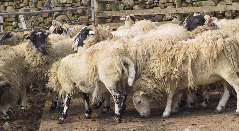
Figure 1. Animals at different stages of disease can be seen in the same flock
During the early stages of sheep scab, infestations are not obvious, and animals often appear clinically normal. It can take 40-50 days after infection before signs are seen. There are low mite numbers and very small, virtually undetectable lesions. The first visible signs can be restlessness, rubbing against fence posts, soiled and stained areas of wool, head tossing and loose tags of fleece.
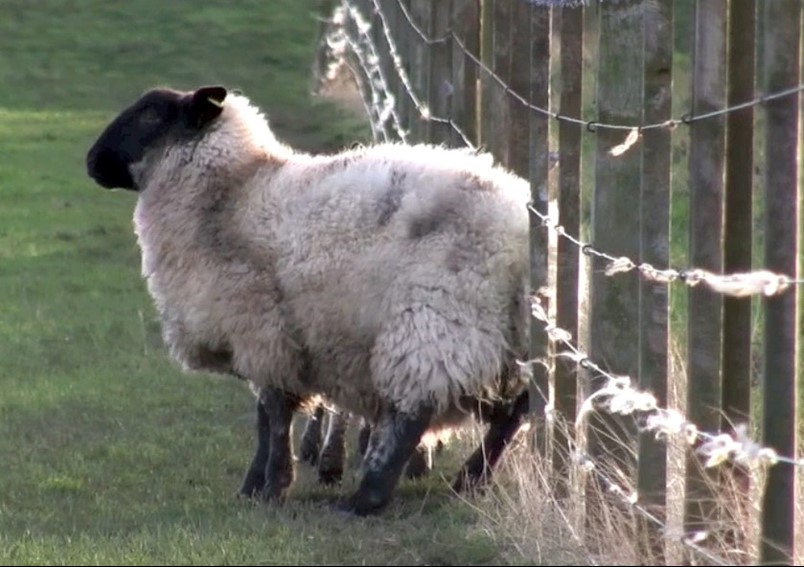
Figure 2. Frequent rubbing against fences and wool tags on the wire may indicate sheep scab or louse infestation
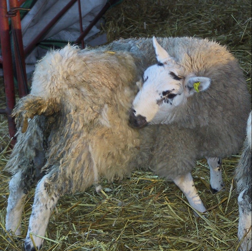
Figure 3. Nibbling the flanks is an early sign of scab and lice
Later stages of infestation with high mite numbers have more obvious clinical signs and the mites spread outwards from the edge of the skin lesions. There is intense rubbing of the shoulders and flanks along the ground or against fences, foot stamping, clawing at the flanks, and biting. Head tossing becomes more excessive and larger areas of wool are lost. There can be open, bleeding wounds and secondary skin infection.
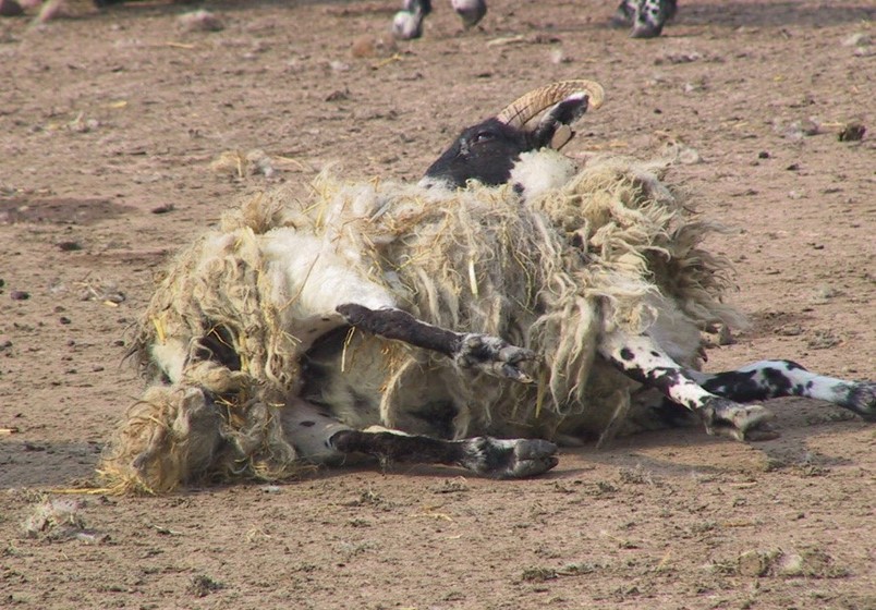
Figure 4. Later stages of sheep scab cause intense itching and biting
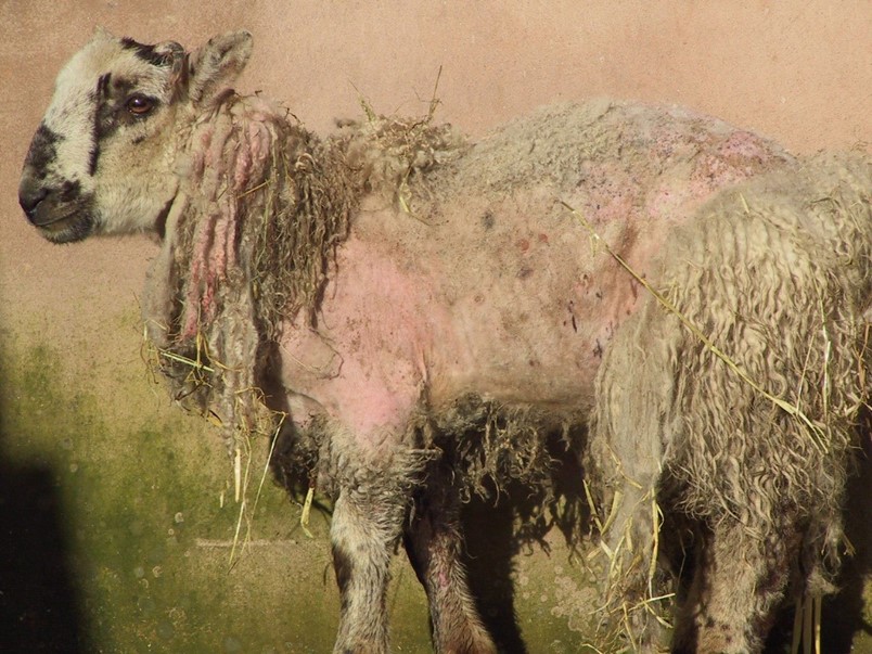
Figure 5. As the disease progresses, wool is lost and the skin becomes thickened.
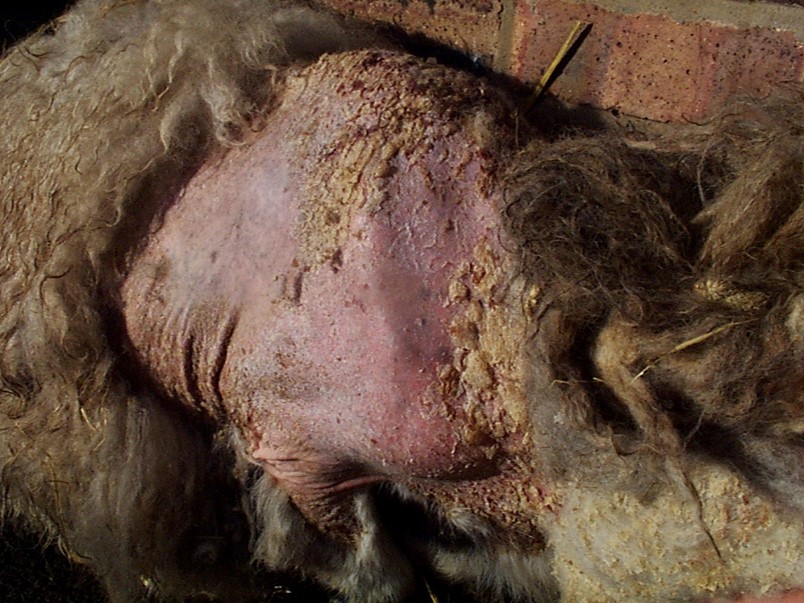
Figure 6. Areas of wool loss with thickened skin become covered with scabs.
The distraction caused by intense itching reduces feeding and there can be a rapid and significant loss in body condition. The loss of fleece and poor nutrition can lead to death through pneumonia or hypothermia. In serious cases, animals will start fitting, particularly when moved.
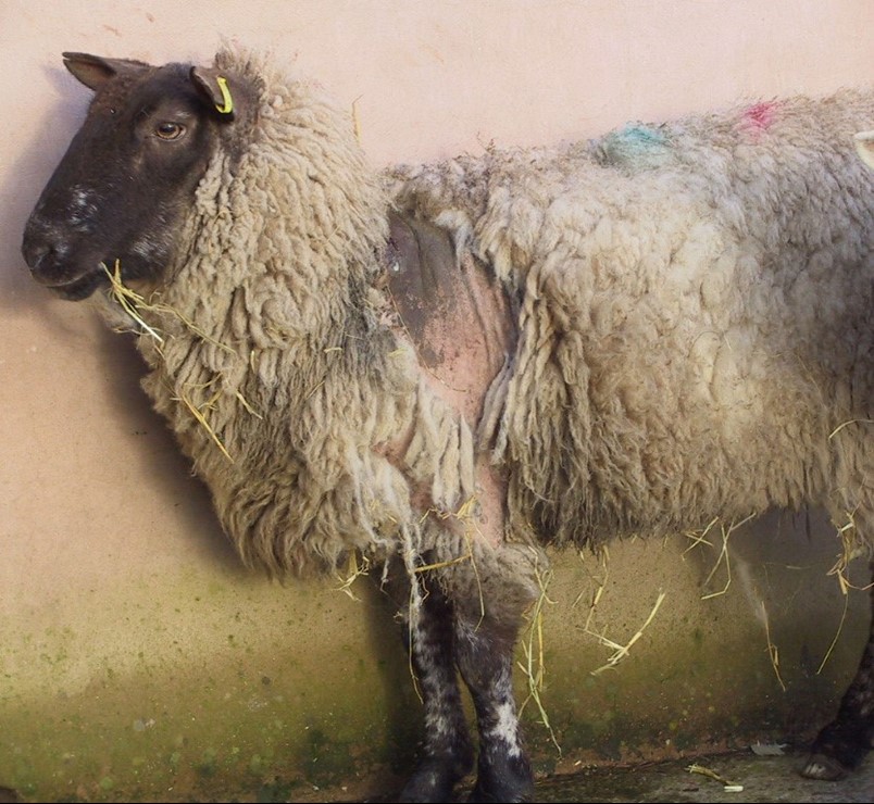
Figure 7. Scab infestation causes rapid weight loss
Low birthweights and higher mortality occur in lambs born to ewes with sheep scab during pregnancy. Slaughterhouse condemnation of carcasses furthers the economic losses.
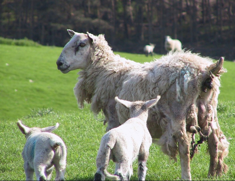
Figure 8: Lower birthweights and higher lamb mortality occurs when ewes have scab
Diagnosis
Clinical signs of sheep scab must be investigated to differentiate them particularly from louse infestation. Falsely attributing the clinical signs to lice contributes to the continued spread of scab. If louse infestations are assumed to be sheep scab treatments are used unnecessarily. Sheep scab and lice can occur together.
Other diseases causing itching include bacterial and fly-bite dermatitis, ked and forage mite infestation, rain scald, lumpy wool and scrapie. Wool loss can occur naturally in some breeds or after housing and when lambs climb on their mothers’ backs. It also occurs after serious illnesses such as mastitis or twin lamb disease and after eating poisonous plants such as St. John’s Wort or rape.
Farmers must seek veterinary advice to get a correct diagnosis. To confirm sheep scab, skin scrapes from the edge of a crusting lesion can be examined under a microscope to identify the mite. This test confirms a current infestation and all sheep in the affected group must be treated. Samples of fleece only or skin scrapes from the centre of a lesion from an infected sheep may not contain mites. Examination of the skin by eye is insufficient. It can confirm a louse infestation but does not rule out the presence of scab mites and both can be present on the same animal.
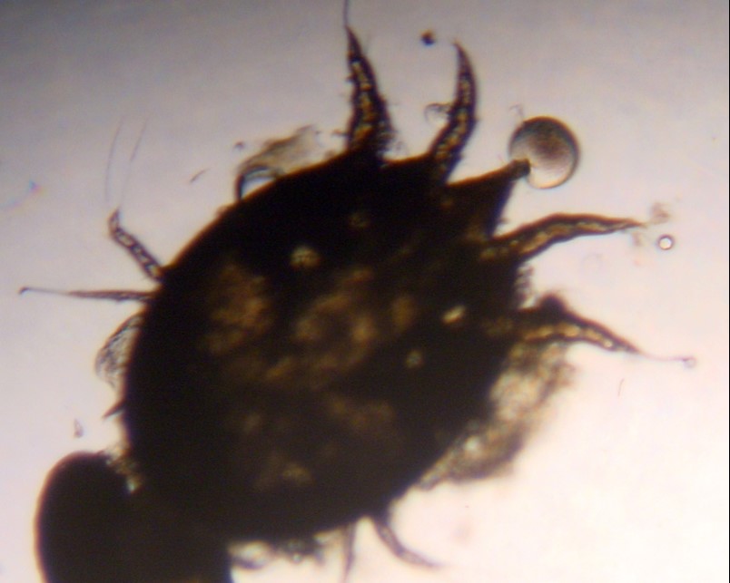
Figure 9. Psoroptes ovis mite under the microscope
In addition to microscopy, sheep can be blood tested for antibodies to sheep scab. The test will not return a positive result for at least two weeks after infection. It is only suitable to test purchased or returning animals which have been isolated for two weeks prior to sampling. Positive results confirm recent exposure to scab mites but the results will remain positive for several months even after effective treatment.
Treatment
There are only two types of treatment available to treat or prevent sheep scab. They are plunge dipping with the organophosphate Diazinon (e.g. Osmonds Gold Fleece Sheep Dip and Paracide 62) or injecting with one of the macrocyclic lactones – moxidectin, doramectin or ivermectin (e.g. Cydectin, Dectomax, Ivomec).
Organophosphate dips are only effective against sheep scab when applied by correctly plunge dipping. They are not effective when used in jets, showers or sprays. Dips kill scab mites within 24 hours and have residual action for several weeks. They provide ongoing protection from re-infection if sheep are returning to the same infected pastures or commons. Dips also control blowfly, lice, keds and ticks.
Operators must have a formal certificate of competence achieved at an approved training course. They must take all the necessary precautions to avoid human and animal health risks and have all the correct permissions or licenses for safe disposal.
Macrocyclic lactones (3-ML) injections when administered correctly can be effective against scab but they do not control blowflies, lice or ticks. The correct dosing procedure for the product must be followed with double dosing for ivermectin and moxidectin 1%. Doramectin and ivermectin have a short residual action and animals are vulnerable to re-infection from other untreated animals or their infected environment (if re-exposed within the 17 day survival period). Sheep treated with these two products may also remain infective for up to 14 days after treatment and must be separated from untreated animals. Moxidectin 2% is injected in the base of the ear and operators must be competent in this technique and aware of the risks.
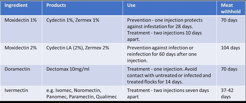
Figure 10. Adapted from SCOPS: https://www.scops.org.uk/workspace/pdfs/scab-product-options-table_1.pdf
All these injectable drugs are very valuable wormers in the 3-ML class and reliance on them for sheep scab control increases the risk of worm resistance developing.
A study published in 2018 confirmed that sheep scab mites resistant to moxidectin had been found in cases in Wales where treatment had failed. This resistance is likely to pass between all the injectables in the group. Any suspicion of treatment failure must be reported to your vet who can confirm the diagnosis, establish the treatment regime used, report the case to the relevant authorities and provide advice on further treatment.
Prevention
The risks of entry of sheep scab can be significantly reduced through good biosecurity. Maintain a closed flock where possible or ensure effective quarantine and treatment for incoming animals. Ensure all boundaries are stock proof and be aware of the risks of entry on people, clothing and equipment.
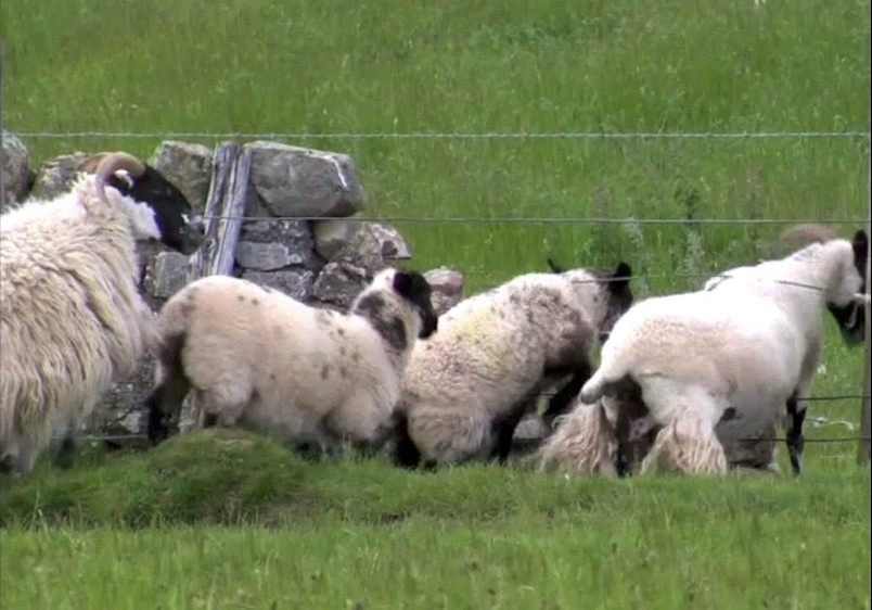
Figure 11. Stray sheep can introduce scab – this boundary is not secure.
All incoming sheep including animals from other farms, markets or off grounds may be carrying scab mites even if they have no clinical signs of disease. They must be assumed as carriers, regardless of any assurances of disease freedom or prior treatment from vendors. Even sheep from scab free flocks can be infected at markets, in transport or from contaminated clothing and equipment.
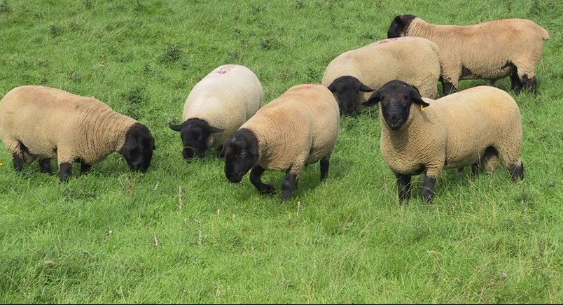
Figure 12. Assume all incoming sheep are infected. Quarantine, test and treat if required.
Farmers must establish and carry out robust biosecurity plans established with their vets as part of the flock health plan. Incoming sheep must be effectively quarantined. The best option to avoid unnecessary scab treatment is to blood test 12 animals in the group for exposure. It is only reliable when samples are collected two weeks after quarantine started.
If blood test results are positive or for untested animals treat the whole group with an effective product according to your health plan and continue to isolate them from the rest of the flock before mixing for the period set out in your health plan. Quarantine options for worms and scab are available from SCOPS: https://www.scops.org.uk/workspace/pdfs/quarantine-options-table.pdf
Common grazers have the highest risk of scab and to gain control all graziers with animals on the same common must cooperate. On established “off-days” it is essential to round up all sheep from the common. This is difficult in practice and some studies estimate 10% of upland sheep can be missed at gathering. Treatment must have residual, preventative action if the animals are returning to the same pasture – OP plunge dip or moxidectin 2%. Animals with short acting treatments could be re-infected from mites in the environment or eggs hatching from other sheep treated similarly. Local authorities have legal powers to ensure animals on affected commons are treated – farmers can report to the local authority if other keepers are failing to control infection.
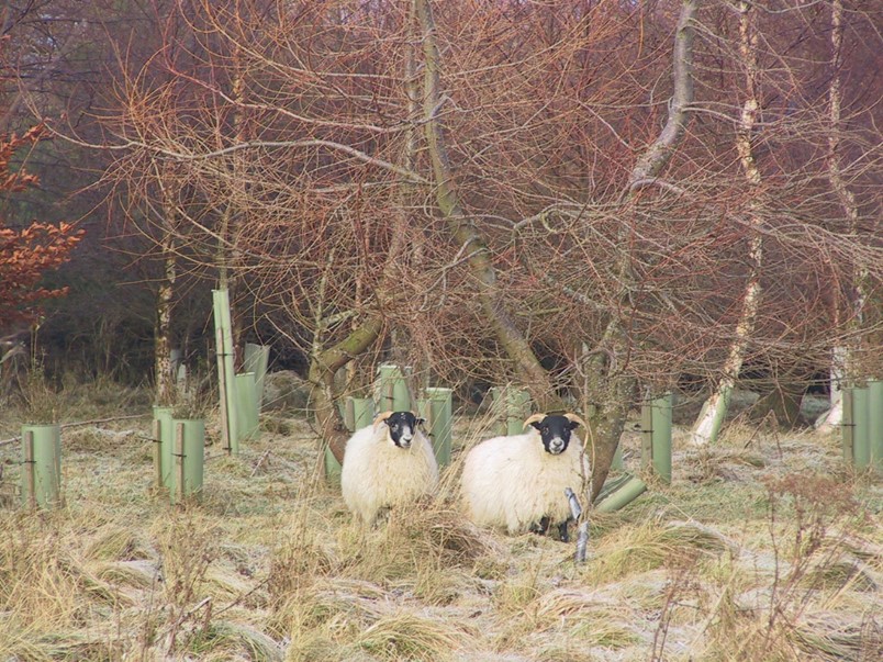
Figure 13. Untreated sheep on commons pose a risk of re-infection. Use products with residual action for sheep returning to common grazing
Subscribe to:
Posts (Atom)

-
Anthrax is a serious infectious disease caused by gram-positive, rod-shaped bacteria known as Bacillus anthracis . It occurs naturally...
-
Lumpy cow skin disease is a concern for cattle owners, leading to significant economic losses worldwide. Thus, many are eager to know wh...


